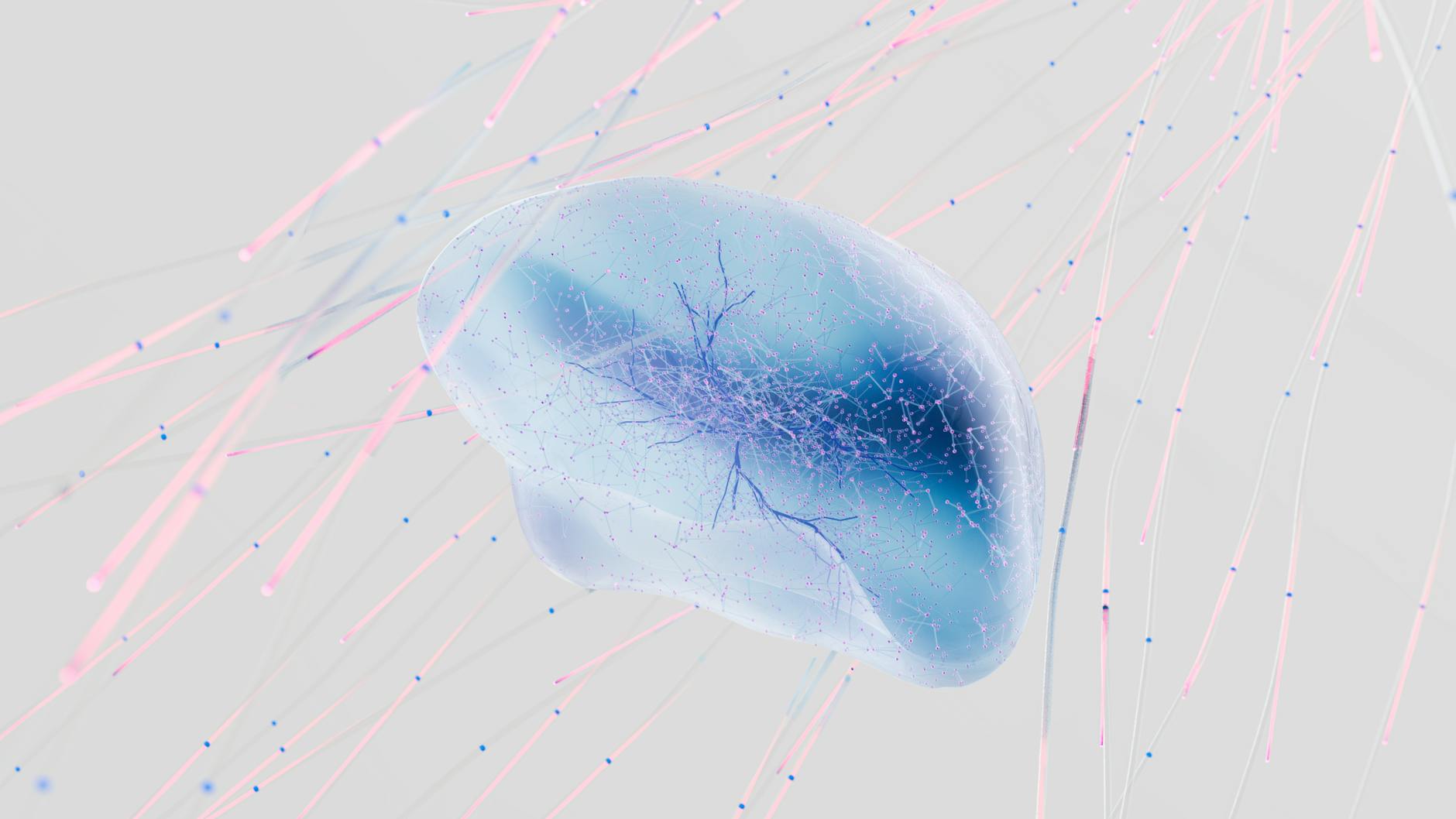
Scientists have pinpointed a specific class of cortical inhibitory neurons—Sst‑Chodl cells—that can trigger sleep when stimulated in mice.
Key Takeaways
- Sst‑Chodl neurons are a distinct population of cortical inhibitory cells.
- When activated, these neurons initiate sleep in mice.
- The finding was made using a mouse model.
- It offers a new angle on how the brain regulates sleep onset.
| Metric | Value | Why it matters |
|---|---|---|
| Cell type | Sst‑Chodl neurons | Key to understanding sleep onset mechanisms. |
| Function | Initiate sleep when activated | Potential target for sleep‑related therapies. |
| Model organism | Mice | Provides a controlled system to study neural circuits. |
What Are Sst‑Chodl Neurons?
Sst‑Chodl neurons belong to the broader family of cortical inhibitory cells, which play a pivotal role in balancing excitatory signals within the brain’s cortex. The “Sst” designation refers to the expression of somatostatin, a neuropeptide that modulates neuronal activity, while “Chodl” indicates the presence of the chondrolectin protein. This combination of markers distinguishes them from other inhibitory subtypes.
How the Discovery Was Made
The identification of Sst‑Chodl neurons as sleep‑inducing cells emerged from targeted experiments in mice. By selectively activating these neurons, researchers observed a consistent onset of sleep behavior. The methodology relied on precise stimulation techniques that can be applied to specific neuronal populations, allowing for clear attribution of the sleep response to Sst‑Chodl activity.
Implications for Sleep Research
Sleep regulation is orchestrated by a complex interplay of neuronal circuits. The discovery that a single inhibitory cell type can initiate sleep adds a new layer to this understanding. It suggests that the brain may employ dedicated “sleep‑switch” neurons that, when engaged, can override other arousal signals. This insight could guide future investigations into how sleep disorders arise when such switches malfunction.
Future Directions
Building on this finding, researchers may explore whether similar neuronal populations exist in other species, including humans. Comparative studies could determine if Sst‑Chodl‑like cells contribute to natural sleep cycles or if they can be harnessed therapeutically. Additionally, mapping the upstream inputs that activate these neurons will clarify how external cues translate into sleep initiation.
Why it Matters
Understanding the cellular underpinnings of sleep is crucial for addressing conditions such as insomnia, hypersomnia, and circadian rhythm disorders. The identification of a sleep‑initiating neuron type opens avenues for developing interventions that target specific neural circuits, potentially offering more precise treatments with fewer side effects than current pharmacological options.
Frequently Asked Questions
What exactly are Sst‑Chodl neurons?
They are a specialized group of cortical inhibitory cells that express both somatostatin and chondrolectin. This unique combination of markers sets them apart from other inhibitory neuron subtypes.
How were these neurons identified as sleep‑inducing?
By selectively activating them in mice and observing a reliable onset of sleep behavior, researchers linked the activity of Sst‑Chodl neurons directly to sleep initiation.
Does this discovery apply to humans?
The study was conducted in mice, so further research is needed to determine whether analogous neurons exist in humans and play a similar role in sleep regulation.
What could this mean for sleep disorder treatments?
If similar mechanisms exist in humans, therapies that modulate the activity of sleep‑initiating neurons might offer targeted ways to correct sleep disturbances with fewer systemic effects.
What are the next steps for this line of research?
Future work will likely focus on identifying the upstream signals that trigger Sst‑Chodl neuron activation and exploring whether these cells can be manipulated safely in more complex organisms.
Bottom Line
The discovery that Sst‑Chodl neurons can initiate sleep when activated in mice marks a significant step toward unraveling the neural circuitry of sleep. While the findings are currently limited to a mouse model, they provide a promising framework for future investigations that could eventually translate into novel, circuit‑specific treatments for sleep disorders.
Reporting based on: original source.
Comments
Post a Comment